Managing dental conditions is not a part of most nursing positions and little dental training is provided in nursing school. However, correctional nurses are most often the first healthcare staff to evaluate a dental complaint and make determinations regarding referrals for treatment and urgency of care delivery. Most intake screenings include a review of dental conditions. In the next few weeks, I will be discussing basic dental and oral anatomy; common dental conditions and appliances; and review some common medical conditions that may affect oral health; topics that are important to correctional nursing practice.
This information is from The Correctional Nurse Educator class entitled Dental Concepts for the Correctional Nurse.
Knowledge of basic oral and dental anatomy is important to gain an understanding of dental evaluation and treatment. Terminology is especially important in the documentation of assessment findings.
Dental Anatomy
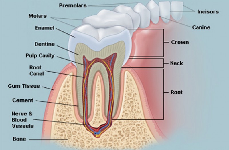
A tooth has two main sections: the crown and the root. The crown sits above the gum line and is covered by enamel. Under the enamel is dentin and in the center of the tooth is the pulp. The pulp holds the nerves and blood vessels that supply the tooth. The root is below the gum line and is covered by cementum. Cementum is softer than enamel.Adults have a total of 32 teeth; 16 in the maxillary row and 16 in the mandibular row. Upper (maxillary) and lower (mandibular) teeth are made up of four types: incisors, canines, premolars, and molars. When determining tooth pain, injury, or condition, it is helpful to document the location and type of tooth. For example: “Severe pain localized to right maxillary canine tooth”. Although the dental profession uses a universal numbering system, unless you are trained and required to use it, it is best to document descriptive locations as in the example above.
Oral Anatomy
Many dental conditions also involve other structures in the mouth, and documenting the location and size is important.
The roof of the mouth consists of a hard palate as a foundation to the maxillary teeth and a soft palate extending back to the oropharynx. The floor of the mouth is a foundation for the mandibular teeth and connects the tongue at the posterior end near the oropharynx. Salivary glands include the parotid, the submandibular (or submaxillary), and the sublingual. The cheeks encircle the mouth interior and are lined with protective mucosa. This mucosa is firmer over the gum area.
In Dental Concepts II, we will discuss Dental Assessment at Intake.